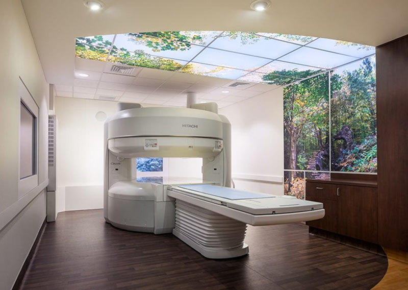
MRI stands for magnetic resonance imaging. It is a diagnostic imaging technique that allows your doctor to look inside your body to diagnose disease and evaluate the effectiveness of treatment. There are benefits and drawbacks to an MRI. Magnetic resonance imaging does not expose you to radiation the way a CT scan or x-ray would. On the other hand, while an x-rays or CT can produce an image of your bones, an MRI only visualizes soft tissues in your body.
What Happens During an MRI Scan?
Usually, an MRI requires you to lie on a table that slides into a cylindrical machine that performs the scan. However, some people experience claustrophobia and have difficulty entering an enclosed space for purposes of the exam. If you have claustrophobic issues, a scan from an open MRI machine may be a good alternative.
In either case, you will have to remain very still while the MRI is in progress. Any movement can show up as an artifact on the final image. Depending on the body part targeted, the process can take anywhere from 10 minutes to an hour or more. During this time, the natural alignment of hydrogen atoms in your body is temporarily altered due to a combination of radio waves and a strong magnetic field that the machine generates around your body. The nuclei of the hydrogen atoms send out radio signals as they align back into proper position. The computer converts these signals into 2D images following analysis.
What Conditions Do MRIs Diagnose?
MRI can be used to detect abnormalities in soft tissues throughout the body. It can be used to detect breast cancer, heart valve problems, torn ligaments, aneurysms, and injuries to the brain or spinal cord. An MRI can help to diagnose stroke and is often instrumental in identifying the damage to the outer protective covering of nerve cells that indicates multiple sclerosis.
Though limited in some ways, and MRI is an essential diagnostic tool. Doctors use MRIs to help patients on a daily basis.


